The authors suggest a technique to improve the accuracy of measuring IOP in upgaze using Goldmann applanation tonometry.
Graves’ ophthalmopathy, also known as thyroid eye disease, is an autoimmune inflammatory condition affecting the orbit and periorbital tissues [1]. It was first described in 1835 by Irish surgeon Robert James Graves [2]. Graves’ ophthalmopathy is commonly characterised by lid retraction, lid lag, swelling, conjunctivitis, keratitis and proptosis [3]. In severe cases, impaired visual acuity and visual field defects can also occur [3].
Graves’ ophthalmopathy commonly causes elevated intraocular pressure (IOP) in upgaze by 1-15mmHg (in 65–100%), which is explained by a thickened and inelastic inferior rectus muscle indenting the globe and blocking episcleral aqueous outflow [4-6]. However, IOP can also increase in upgaze in normal subjects [4,7]. Hence, elevated IOP in upgaze can be considered a normal phenomenon exaggerated by Graves’ ophthalmopathy.
The Goldmann applanation tonometer (Haag-Streit, Wedel, Germany) provides gold standard IOP measurements [8]. It is mounted on the slit-lamp which keeps the patient’s chin and forehead in a fixed position. The chin rest is adjusted until the lateral canthus is in line with the eye level marker, such that the tonometer prism is lined up with the central cornea in primary gaze. This maintains consistency and accuracy in IOP measurement in primary gaze. However, when the patient is asked to look upwards, the inferior limbus is applanated rather than the central cornea [9]. This may artificially increase the IOP reading, potentially producing false positives in those assessed for Graves’ ophthalmopathy. Handheld IOP measuring devices such as the Tonopen [9] and Icare [10] may be positioned against the central cornea in upgaze, but these devices are not considered to achieve the same accuracy in IOP measurement as compared to the Goldmann applanation tonometer, nor do they enable consistency in the site and angle of contact with the central cornea.
“Elevated IOP using this technique should provide more compelling evidence of active thyroid eye disease”
Herein, we present a standardised technique to improve accuracy of measuring intraocular pressure in upgaze using gold standard Goldmann applanation tonometry.
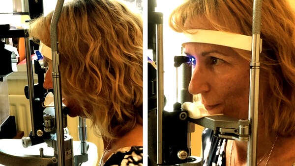
Figure 1: Forward-tilt technique. Left: The patient’s mentolabial sulcus (groove between lower lip and chin) is positioned onto the chin rest. Right: The chin rest is raised until the lateral canthus lines up with the eye level marker (*). The head is tilted forward until it is fixed against the headrest. Whilst in upgaze, the central cornea is applanated by the Goldmann applanation tonometer.
Technique
The forward-tilt technique is illustrated in Figure 1 and the practical steps are outlined below.
Step 1
Measure the patient’s IOP in primary gaze using the Goldmann applanation tonometer in the standard fashion.
Step 2
Position the patient’s mentolabial sulcus onto the chin rest. The mentolabial sulcus is the groove between the lower lip and the chin. See Figure 1A.
Step 3
Raise the chin rest until the lateral canthus lines up with the eye level marker. See Figure 1B.
Step 4
Ask the patient to tilt their head forward until their forehead rests against the headrest.
Step 5
Applanate the central cornea, thereby measuring IOP in upgaze. Compare with IOP in primary gaze to determine whether the IOP is elevated in upgaze.
Discussion
The current, widely performed method is unstandardised in that the patient may look upwards by different amounts per visit, possibly leading to false assumptions regarding disease activity. By contrast, the forward-tilt technique offers a more standardised approach to measuring IOP in upgaze. Perpendicularity is maintained between the central cornea and prism head whilst the eye is fixed in upgaze, thereby producing more accurate IOP readings as compared to applanation of the inferior limbus. The critical modification of the standard technique is the repositioning of the mentolabial sulcus onto the chin rest, followed by raising the chin rest such that the lateral canthus lines up with the eye level marker, thereby forcing the head into a forward-tilt for applanation of the central cornea with the eye forced into upgaze.
Additional time is required to adjust the height of the chin rest in the forward-tilt technique, but this is minimal. Elevated IOP using this technique should provide more compelling evidence of active thyroid eye disease.
Full clinical workup should be performed for all patients with suspected Graves’ ophthalmopathy in the ophthalmology clinic. This typically includes visual acuity, colour vision, comprehensive ophthalmic examination, visual field testing and orthoptic examination.
References
1.Bahn RS. Graves’ ophthalmopathy. N Engl J Med 2010;362(8):726-38.
2. Graves R. Newly observed affection of the thyroid: clinical lectures. Lond Med Surg J 1835;7:516-7.
3. Char D H. Eye signs and diagnosis. Rootman J. Diseases of the Orbit. Philadelphia: JB Lippincot; 1988:241-80.
4. Spierer A, Eisenstein Z. The role of increased intraocular pressure on upgaze in the assessment of Graves ophthalmopathy. Ophthalmology 1991;98(10):1491-4.
5. Gamblin GT, Harper DG, Galentine P, Buck DR, Chernow B, Eil C et al. Prevalence of increased intraocular pressure in Graves’ disease--evidence of frequent subclinical ophthalmopathy. N Engl J Med 1983;308(8):420-4.
6. Fishman DR, Benes SC. Upgaze intraocular pressure changes and strabismus in Graves’ ophthalmopathy. J Clin Neuroophthalmol 1991;11(3):162-5.
7. Reader AL. Normal Variations of Intraocular Pressure on Vertical Gaze. Ophthalmology. 1982;89(9):1084-7.
8. De Moraes CGV, Prata TS, Liebmann J, Ritch R. Modalities of tonometry and their accuracy with respect to corneal thickness and irregularities. J Optom. 2008;1(2):3-9.
9. Rahman I, Cannon PS, Sadiq SA. Tonopen versus Goldmann applanation tonometry for detecting restrictive thyroid eye disease. Ophthal Plast Reconstr Surg 2010;26(1):6‑8.
10. Rödter TH, Knippschild S, Baulig C, Krummenauer F. Meta-analysis of the concordance of Icare® PRO-based rebound and Goldmann applanation tonometry in glaucoma patients. Eur J Ophthalmol August 2019:1120672119866067.
TAKE HOME MESSAGE
-
Graves’ ophthalmopathy is commonly associated with elevated IOP in upgaze.
-
Strictly speaking, the current technique for IOP measurement in upgaze is unstandardised.
-
The forward-tilt technique offers a more standardised approach to measure IOP in upgaze using Goldmann applanation tonometry.
COMMENTS ARE WELCOME