Congenital and paediatric cataracts are relatively rare, although prevalence varies significantly between countries, influenced by factors such as nutrition, immunisation policy and population genetics [1]. In the UK, around 3–4 of every 10,000 babies are born with cataracts.
It’s a treatable cause of potential visual impairment in children. Despite this, congenital and infantile cataract remain significant causes of lifelong visual impairment. They account for approximately 20% of blindness in children worldwide, and 3% in the UK [1]. This stark difference is explained, at least partly, by access to diagnostic examinations and prompt surgical care. Over the last 40 years, advances in both diagnosis and surgical management of congenital and paediatric cataracts have led to improved visual outcomes. We discuss these advances below:

Figure 1 : Congenital lamellar cataract.
1. Diagnosis – early detection
In the UK, the neonatal and infant physical screening examination (NIPE) includes an eye component, specifically an assessment of the pupillary red reflex [2]. This should be performed within 72 hours of birth and again at 6–8 weeks. Its aim is to detect opacities within the visual axis and thus identify congenital cataracts. When a lens opacity is suspected at the newborn examination, more detailed assessment by an ophthalmologist should be performed within two weeks. If a congenital cataract is suspected later at the 6–8-week examination, urgent referral is indicated, with the recommendation of review by an ophthalmologist by 11 weeks of age. However, this timeframe leaves affected infants at the very limit of the critical period for optimal outcomes, and we recommend that the referral should be made as soon as possible.
NIPE has an uptake of >95% in the UK. This results in early diagnosis for most affected infants, enabling prompt management and thus limitation of sight impairment caused by visual deprivation amblyopia. However, the eye component of the NIPE screening programme is not fail-safe and improved screening instruments, particularly for use by non-ophthalmologists, are currently being developed and validated [3].
Pre-school vision screening is also performed in the UK when children are aged between four and five years. The aim is to detect reduced vision in one or both eyes. When a child fails screening at this age, the cause is typically amblyopia due to refractive error or strabismus. However, a small number of children with cataract are also identified.
Advances in obstetric ultrasound quality mean that it can, on occasions, be possible to image congenital cataracts prenatally. This is particularly useful in pregnant women with a family history of congenital cataract, thus expediting referral for precise diagnosis soon after birth and optimising management [4].

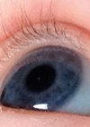
Figure 2 : Congenital posterior congenital cataract with cortical flecks.
2. Advanced imaging and diagnostic modalities
B-scan ultrasound, ultrasound biomicroscopy, electrodiagnostic testing and anterior segment OCT can provide additional useful clinical information to aid surgical planning without the need for an additional examination under anaesthesia.
3. Aetiology
Paediatric cataract is a clinical sign and not a specific diagnosis. There is a plethora of causes which include genetic, metabolic, infectious and traumatic disorders. Genetic aetiology (both eye / lens specific and associated with systemic / syndromic disease) underpins most bilateral paediatric cataract in the developed world [5].
Unilateral congenital cataract is mostly sporadic. It’s often associated with a persistent foetal vascular anomaly or posterior lenticonus of the lens. Infectious causes of cataract are rare in the UK compared to developing nations. These can include toxoplasmosis, rubella, cytomegalovirus, varicella and syphilis. Access to public health initiatives, including vaccination programmes for conditions such as rubella, significantly influence incidence.
Genetic testing
In recent years the development of high throughput, next-generation DNA sequencing has transformed the utility of genomic investigations across all fields of medicine. Genetic testing, enabling precise diagnosis, has now become mainstream in tertiary paediatric cataract clinics. This typically involves trio whole genome sequencing (DNA taken from both parents and the affected child) and initial interrogation of a panel of genes in which mutations are known to cause cataracts. Testing is delivered in combination with careful clinical phenotyping, and involvement of a multidisciplinary team for variant interpretation. Working with a skilled genetic counsellor is essential.
This approach is used in cases of bilateral cataracts and in some children with unilateral cataract children and other systemic abnormalities / dysmorphic features. A causative genetic mutation is identified in most cases of bilateral cataract. Autosomal dominant inheritance is most common [6].
In one UK series, 15% of children with bilateral congenital cataracts were found to have associated systemic issues [6] – the incidence is probably higher in specialist children’s hospitals [7]. Genetic testing can help identify children in whom systemic features are initially subtle or not yet apparent. This can enable early therapeutic input and potential life-changing disease modification in metabolic conditions such as cerebro-tendo-xanthomatosis [7].
Surgical innovations
Until the 1980s, surgery for unilateral cataract was almost universally associated with poor outcomes. However, in a seminal 1981 paper, Beller, et al. reported good visual function in eight infants who had undergone surgery before six weeks of age [8]. Their management included prompt contact lens correction and aggressive occlusion therapy. Birch, et al. further clarified this “critical period” for unilateral cases undergoing surgery [9]. Early surgical intervention for unilateral cataracts, when referred promptly, has since become the norm. This includes those associated with mild- to moderate-anterior persistent foetal vasculature, a common association of unilateral congenital cataract.
Two well-constructed studies (IATS and IOLu2) both concluded that implantation of IOLs is not recommended for young infants, particularly where access is available to a good paediatric contact lens service [10,11]. Both studies found no difference in acuity outcomes between infants corrected with a contact lens compared to those with an IOL. However, the re-operation rate in the IOL groups was noted to be much higher, largely due to visual axis opacification. This led to the recommendation that IOL implantation is limited to infants at risk of significant periods of uncorrected high refractive error if left aphakic. This would include families likely to have difficulty with managing a contact lens, those with a developmentally delayed child or those living very far from the hospital. A major advantage of contact lens correction over IOL implantation is that changes in refraction can be easily corrected by modifying the lens strength rather than relying on additional spectacles.
In infants and young children undergoing cataract surgery, visual axis opacification (PCO) is rapid and inevitable if the posterior capsule is left intact. Thus, primary posterior capsulectomy and vitrectomy are recommended by most surgeons in children under the age of five years [2]. In older children, manual posterior capsulotomy can be performed without a vitrectomy. Posterior optic capture may additionally be performed to limit cellular migration of cells across the visual axis.
Single-piece hydrophobic acrylic IOLs are commonly used in older children. Three-piece acrylic IOLs, with posterior angulation designed to reduce pupillary capture and provide contact inhibition to limit / delay PCO, are suitable for either sulcus fixation or in-the-bag positioning. They are the lens of choice when performing optic capture.

Figure 3 : Dense unilateral congenital cataract associated with persistent fetal vascular malformation.
4. Timing of surgery in infants
Development of the infant visual system is profoundly affected by visual deprivation. Prolonged deprivation causes irreversible changes in both the lateral geniculate nuclei and the visual cortex. However, very early surgery, particularly in the first month, is associated with an increased risk of aphakic glaucoma [12]. Thus, timing of surgery must balance this risk of secondary glaucoma with the risk of undue delay causing visual deprivation.
There is some evidence that in very early neonatal life, the immature visual system relies on sub-cortical pathways. During this sub-cortical or latent period, transient visual disturbance does not appear to impact substantially on eventual visual outcome. This period appears to be approximately six weeks for human infants with unilateral visual deprivation and postulated to be as long as 10 weeks in bilateral visual deprivation [13]. However, the IoLu2 study concluded that, in the first three months, each additional month at cataract surgery led to a progressively worse visual outcome [11]. Thus, most surgeons aim to schedule surgery at around six weeks in unilateral cases and eight weeks in bilaterals.
Postoperative management
Postoperative anti-inflammatory treatment using steroid / antibiotic drops and cycloplegia is important and should be monitored carefully. This may need to be aggressive, particularly where there has been perioperative significant iris manipulation due to microcoria or other developmental anomalies.
Early, accurate and regular refractive correction, via aphakic glasses, and / or contact lenses is essential. Occlusion therapy is a very important part of postoperative treatment in unilateral paediatric cataracts and is key to good outcomes. This should be guided by regular orthoptic assessment and acuity monitoring.
Conclusion
Optimal management of congenital and paediatric cataract in the current era is multidisciplinary, technology assisted and patient centred. Early and precise diagnosis, modern surgical techniques, customised IOL use, and robust amblyopia therapy improve visual outcomes.
Further advances are likely to involve genetic research, in combination with deep phenotyping, advanced imaging and neurophysiology. Novel therapies linked to metabolomics may also play a role. Early detection, appropriately timed and careful surgery, and rigorous postoperative management currently remain the key factors in the optimisation of visual outcomes.
References
1. Wu X, Long E, Lin H, Liu Y. Prevalence and epidemiological characteristics of congenital cataract: a systematic review and meta-analysis. Sci Rep 2016;6:28564.
2. Self JE, Taylor R, Solebo AL, et al. Cataract management in children: a review of the literature and current practice across five large UK centres. Eye (Lond) 2020;34(12):2197–218.
3. Duret A, Humphries R, Ramanujam S, et al. The infrared reflex: a potential new method for congenital cataract screening. Eye (Lond) 2019;33(12):1865–70.
4. https://www.fetalultrasound.com/online/text/30-137.HTM
5. Musleh M, Hall G, Lloyd IC, et al. Diagnosing the cause of bilateral paediatric cataracts: comparison of standard testing with a next-generation sequencing approach. Eye (Lond) 2016;30(9):1175–81.
6. Bell SJ, Oluonye N, Harding P, Moosajee M. Congenital cataract: a guide to genetic and clinical management. Ther Adv Rare Dis 2020;1:2633004020938061.
7. Gillespie RL, Urquhart J, Anderson B, et al. Next-generation Sequencing in the Diagnosis of Metabolic Disease Marked by Pediatric Cataract. Ophthalmology 2016;123(1):217–20.
8. Beller R, Hoyt CS, Marg E, Odom JV. Good visual function after neonatal surgery for congenital monocular cataracts. Am J Ophthalmol 1981:91(5):559–65.
9. Birch EE, Stager DR. The critical period for surgical treatment of dense congenital unilateral cataract. Invest Ophthalmol Vis Sci 1996;37(8):1532–8.
10. Infant Aphakia Treatment Study G. Lambert SR, Lynn MJ, et al. Comparison of contact lens and intraocular lens correction of monocular aphakia during infancy: a randomized clinical trial of HOTV optotype acuity at age 4.5 years and clinical findings at age 5 years. JAMA Ophthalmol 2014;132(6):676–82.
11. Solebo AL, Russell-Eggitt I, Cumberland PM, et al. Risks and outcomes associated with primary intraocular lens implantation in children under 2 years of age: the IoLunder2 cohort study. Br J Ophthalmol 2015;99(11):1471–6.
12. Trivedi RH, Wilson Jr ME, Golub RL. Incidence and risk factors for glaucoma after pediatric cataract surgery with and without intraocular lens implantation. J AAPOS 2006;10:117–23.
13. Lambert SR, Lynn MJ, Reeves R, et al. Is there a latent period for the treatment of children with dense bilateral congenital cataracts? J AAPOS 2006;10:30–6.