We hope you like and appreciate the image forming the cover of the June/July 2021 edition of Eye News. The Ophthalmic Imaging Association (OIA) was honoured to have been invited to submit a series of images from the winners of our annual Imaging Competition, to present as cover-page material for the next few editions.
OIA was formed over 30 years ago, as a ‘professional learned society’, by a group of ophthalmologists and ophthalmic photographers. Key persons driving the establishment of the first meeting were Drs Bill Haining and Emanuel Rosen and, of course, recently retired Head of Imaging at Moorfields, Kulwant Sehmi, along with other ‘big names’ of the profession.
The initial ‘fact-finding’ meeting, held in Harrogate, saw over 100 professionals meet to lay out the plan for an association dedicated to share best-practice, nurture new-comers to the profession and, critically, to help to improve ocular imaging to the ultimate benefit of patients. The symbiosis between ophthalmologists with keen interests in ophthalmic imaging and specialist ophthalmic or general medical photographers with an interest in ophthalmics was crucial to success and remains so to this day. When formed, our association was actually called the British Ophthalmic Photographic Association, or BOPA.
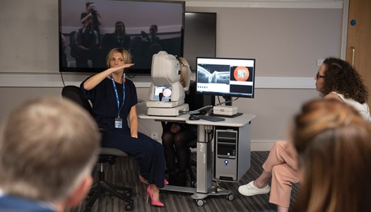
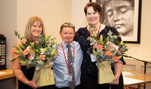
Photos taken at the OIA Annual Conference 2019.
Several years later, to recognise that we had interest from professional staff all over the world, we changed our style from BOPA to become OIA. Two important changes there – removal of ‘British’ (although we are still grounded in the UK and the bulk of members remain UK-based), and Photographic becoming Imaging to reflect the changes in technology and terminology.
From the outset, the aims of BOPA / OIA were to support best practice in ophthalmic imaging. Our remit remains the same: “Promoting good practice, education, training and research within the discipline of ocular imaging.” More information can be found here: https://eyeimaging.org/
The development of ophthalmic imaging
In the early days of the Association, all our recording of ocular features was of course carried out using film. Those days will be fondly (?) remembered, when we waited with great anticipation to find out if our Kodachrome films, costing about £5.00 each, processed only by Kodak itself and returned in the post some FIVE days later, actually captured what we hoped when we had our patient contact. Of course, we also routinely used other colour transparency (slide) films, usually E6-processed more locally by professional processing labs including Argentum Labs, staffed by ex-ophthalmic photographers (so they knew our needs and requirements).
“We waited with great anticipation to find out if our Kodachrome films actually captured what we hoped when we had our patient contact”
We got those films back to review within a day or so and most of us had the benefit of processing our black-and-white negative films (for fluoresceins) and graphic artwork material ourselves in our own darkrooms. Oh, the smells associated with acetic acid ‘stop-bath’, ammonia-processed diazos and the joys of deep-tank processing up to 36 rolls of 35mm fundus fluorescein angiography (FFA) film at a time under weird Kodak 1A safelight conditions.
By the turn of the millennium, most departments started to experiment with the new kid on the block – digital imaging. It was VERY expensive and, to be brutally honest, not very good. It has taken nearly 20 years for digital imaging systems to develop to the point that they are at least as good as the film-based recording media we used in the ‘good old days’. Excellent digital imaging systems are now much more affordable and have running costs (‘consumables’) close to zero. With digital imaging, we can see pretty much exactly what we have recorded in real-time. No more cases of the 35mm film not going through the camera back properly, no more failures in film processing, dust and water marks and making multiple copies of images is now so simple.
We have now got many more imaging options available to us than we had when I started almost 40 years ago. Who could now conceive of a modern ophthalmic imaging and diagnostics suite without access to optical coherence tomography (OCT), optical coherence tomography angiography (OCT-A), indocyanine green (ICG), microperimetry, etc.? With those additions to our imaging and diagnostic arsenal comes, of course, the need to widen the skill-set of the staff providing the service. New devices and techniques = new opportunities for wonderful diagnostic images.
The imaging competition
This brief review of our history of ophthalmic imaging technology is mirrored in the course of the professional imaging competition which BOPA / OIA have run pretty much every year since inception of our association. Timed to coincide with our Annual Professional Conference each November, we invite members to submit their best work to be judged by a panel of three judges from their senior peers. Entries are (now) judged prior to or (previously) during the Conference, with prize-winners announced during our legendary Friday night conference dinner. Rather like the Oscars, but without the tears and tedious “I would just like thank you Mum, my agent and all my dogs…” speeches.
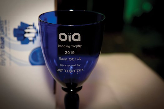
The competition currently has seven technical categories into which members can submit their images: Posterior Segment, Anterior Segment, FFA / ICG Angiography, OCT, OCT-A, Ophthalmic Related and Case Report. Each category has a first and second-placed winner and from all the first-place winners, an overall competition champion is selected by the three independent old people who make up that year’s judging panel assessing all entries. Every category winner receives a beautiful engraved Bristol Blue goblet (see image) and cash-equivalent voucher to use against future OIA courses or conference costs. Runners-up in each category also receive a voucher in recognition of their ‘so very nearly the winner’ submissions.
Most of us like to enter competitions from time to time, but the annual imaging competition leaves nothing to chance. It is a competition centred purely on the skill and professionalism of the members submitting their images. The desire to be a category winner is understandably fierce. To achieve the ‘best of the best’ recognition from those category winners is, of course, the ultimate accolade that year. In recognition of this, our annual competition has had a truly exceptional first prize: an all-expenses-paid trip to attend either the next US-based Ophthalmic Photographers’ Society (OPS) annual professional meeting in the States or, if coinciding with an up-coming quadrennial International Conference on Ophthalmic Photography (ICOP), a trip there, wherever it may be in the world. Nothing compares.
“We have now got many more imaging options available to us than we had when I started almost 40 years ago”
None of these prizes and awards would be possible without the generous and outstanding support of our competition sponsor and OIA Trade Members. TOPCON (Great Britain) Medical Limited have been sponsoring our annual professional imaging competition now for several years, having taken over that sponsorship role from Carl Zeiss Meditec UK, who generously supported the competition from inception. TOPCON have been stalwart supporters of OIA for many years as one of our bunch of wonderful Trade Members. Without them, our Association could not continue. Special mention has to go to TOPCON ‘rep’ Richard Ball, who has served on the OIA Committee for many years, having been re-elected several times by his peers from the Trade Members. We recently recognised Richard’s outstanding contribution and support of OIA, particularly in managing difficult negotiations for the continued support for the sponsorship of our imaging competition, when he was awarded Honorary Fellowship and Life Membership of OIA.
So, in summary, please enjoy this and subsequent cover images of the journal and marvel at the skill and professionalism shown by the expert imagers who submit their material for the annual competition. There is nothing lucky about being a category or overall winner. Only the best will achieve such recognition as judged ‘blind’ by the team of senior peers. The image definitely does not have to be of a rare or unusual clinical case – it is the skill and professional approach that is being judged, not access to esoteric clinical conditions (although we learn much from those, when submitted as well of course!).
OIA welcomes warmly new members who have a passion for ophthalmic imaging, not just those who have chosen to make it their professional career. We want and need new members, be they professional imagers, ophthalmologists (human or veterinarian!), optometrists, ophthalmic nurses or anyone who can share in the journey to constantly improve ocular imaging to the overall benefit of patient care.
![]()
https://eyeimaging.org
COMMENTS ARE WELCOME