Cataract has been documented to be the most significant cause of bilateral blindness in India, where vision <20/200 in the better eye on presentation is defined as blindness [1,2]. Estimation of blindness in India by the World Health Organization (WHO) show 11.2% of the population suffering from preventable blindness, with more than two million new cases expected each year [3].
Nearly 70% of India’s population resides in rural areas, with only 10,000 ophthalmologists responsible for the care of the entire population – a ratio of one ophthalmologist per 100,000 people. Cataract surgery in India poses additional challenges due to lack of modern phacoemulsification equipment and advanced pathology encountered in the eye clinic. Manual small-incision cataract surgery (MSICS) is a low-cost, small-incision form of extracapsular cataract extraction (ECCE) that is principally employed in the developing world. Compared to traditional ECCE, MSICS has the advantage of a self-sealing sutureless wound. In resource poor settings MSICS also has several distinct advantages over phacoemulsification, including shorter operative time, less need for expensive technology and lower overall cost overheads [4,5,6,7]. Recent investigations have shown comparable outcomes and complication rates between patients undergoing phacoemulsification and MSICS with posterior chamber intraocular lens placement [8,9].
Akhand Jyoti Eye Hospital
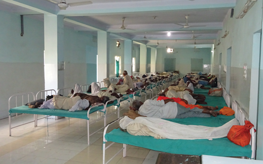
Akhand Jyoti runs a highly successful eye hospital and rural outreach prevention program that aims to reduce the backlog of surgeries to cure blindness in Bihar by 75% in the next five years. The 350-bedded hospital offers primary and secondary level of eye care, including full eye screening, prescription and supply of low cost spectacles, diagnostic evaluation, cataract surgery and other minor surgical procedures. The hospital’s financial model combines income from fee-paying patients with grant subsidy. It aims to provide 2/3 of all services for free and conducts just under 55,000 ophthalmic surgeries a year. Outreach is the core activity of the organisation. It comprises of camps, village door to door activities and school screening. The hospital has a 55 strong outreach team and a large volunteer base.
My journey
One of the reasons for choosing ophthalmology as a career choice, was the scope for volunteering in a ‘vision aid’ project and doing my part to bring the gift of sight to those who would, otherwise, be condemned to be without it. As a visitor to the hospital, my own particular role consisted of providing teaching to the first and second year optometry students. The day began at 7am with breakfast in the hospital canteen, before the daunting task of lecturing 30 optometry students on anatomy, pathology and basic clinical ophthalmology for over an hour. One of the most striking features of the class was the over-whelming female preponderance.
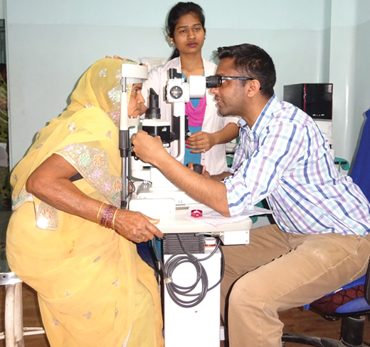
Akhand Jyoti has focused on girls from at-risk communities and runs a parallel education and empowerment program for girls who will eventually have the opportunity to be trained and employed as skilled nurses and optometrists. After class, I was able to go into the main hospital complex and assist in clinics, which run until lunchtime. The running of outpatient department is not too different from our own European set-up – with specialised clinics for glaucoma running on Mondays and Wednesdays, cornea on Tuesdays and medical / surgical retina on Thursdays. A visiting paediatric ophthalmologist would attend on a monthly basis. Patient load is onerous and an ophthalmologist can expect to see anywhere between 40-70 patients in one sitting. Refraction clinics run all day and an onsite dispensing optician is available to provide spectacles.
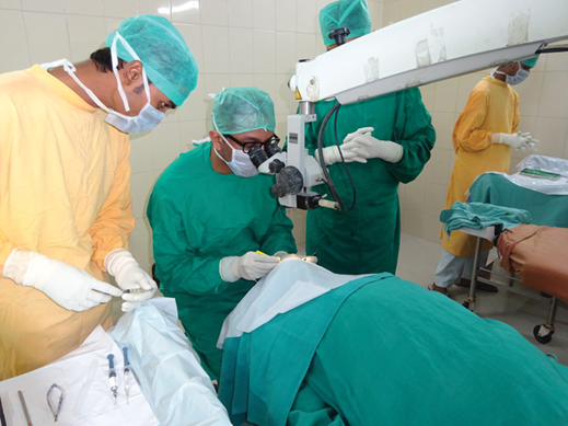
Curiously no specific eye casualty clinic was to be seen – patients with ocular emergencies such as retinal detachment or angle closure were generally treated as and when they presented to the hospital. Afternoon clinics run from the end of lunchtime until around 6pm in the evening, but it was not uncommon to see preoperative sessions going on well into the night. Theatre sessions run throughout the day in a dedicated operating suite. Each patient for surgery is given a retrobulbar block and waits patiently in a queue. Surgical time for each case is at a phenomenally fast rate (average time of seven minutes per case). It is heartening to watch the amazing surgeons at work, who help to complete around 250-300 MSICS procedures a day.
The overwhelming pathology treated in Akhand Jyoti is cataract. Patients are sourced from nearby villages by 50 outreach staff and 700+ volunteers, otherwise known as the ‘guardians of sight’. These guardians of sight work in areas of 10 square kilometres – ensuring that they go to each house with a 3 metre tape and measuring the visual acuities of whoever they encounter. Patients with hand movements or counting fingers are asked to report to the nearby eye screening camp of the hospital. The vast majority of cataracts are mature / hyper mature lens opacities, in which fundal examination is not possible. Biometry is carried out with a keratometer and A-scan, with rigid PMMA intraocular lens widely in use.
The hospital caters to fee paying patients, who can upgrade to toric / multifocal lenses and pay for their surgery to be done by phacoemulsification. In return for my assistance in teaching and clinic work, I was allowed to attend theatres daily, where the medical director allocated me to a resident to learn MSICS. Prior preparation on my part consisted of using simulated eye models to practise the steps of the surgery and reviewing online videos. The most technically difficult step of the procedure is the creation of the scleral tunnel. Too deep and one can risk creating a button hole with the iris, too shallow and the risk of early amputation of flap or non-sealing of wounds poses a real problem. Continuous curvilinear capsulorrhexis size must be larger than the standard 5mm for the polymethyl methacrylate (PMMS) lens to be fitted into the bag. Cataract extraction is either with a fishhook (created with a 30G needle) or irrigating vectis. The wounds created are self-sealing and sutureless and closure is with light conjunctival cautery.
Conclusion
My visit to this remarkable institution impressed on me how important global ophthalmology is. This is really something all ophthalmic trainees can get involved in and the experience is both humbling and edifying to say the least. The treatment of cataract is a life-changing experience for the patients of Akhand Jyoti – many of whom would otherwise not be able to carry out their activities of daily living, go to school / work or interact with their families. The skills learned from MSICS are transferable and reproducible in a teaching hospital setting in the United Kingdom [10]. I would encourage any trainee to try and learn this skill, as potential back-up for the cataract not amenable to routine phacoemulsification. Akhand Jyoti is certainly a beacon of light in a world of darkness and I intend to return there in the future.
References
1. Thulasiraj RD, Nirmalan PK, Ramakrishnan R, et al. Blindness and Vision Impairment in a Rural South Indian Population: The Aravind Comprehensive Eye Survey. Ophthalmology 2003;110:1491-8.
2. Nirmalan PK, Thulasiraj RD, Maneksha V, et al. A population based eye survey of older adults in Tirunelveli district of south India: Blindness, cataract surgery and visual outcomes. Br J Ophthalmol 2002;86:505-12.
3. Dua AS, Muralikrishnan R, Praveen RK, et al. NCMH Background papers; Burden of disease in India. 2005;299-305.
4. Kongsap P. Visual outcome of manual small-incision cataract surgery: comparison of modified Blumenthal and Ruit techniques. Int J Ophthalmol 2011;4(1):62-5.
5. Ruit S, Tabin GC, Nissman SA, et al. Low-cost high-volume extracapsular cataract extraction with posterior-chamber intraocular lens implantation in Nepal. Ophthalmol 1999;106(10):1887-92.
6. Ruit S, Paudyal G, Gurung R, et al. An innovation in developing world cataract surgery: sutureless extracapsular cataract extraction with intraocular lens implantation. Clin Experiment Ophthalmol 2000;28(4):274-9.
7. Blumenthal M, Ashkenazi I, Assia E, Cahane M. Ophthalmic Surg 1992;23(10):699-701.
8. Ruit S, Tabin G, Chang D, et al. A prospective randomized clinical trial of phacoemulsification vs manual sutureless small-incision cataract surgery in Nepal. Am J Ophthalmol 2007;143:32-8.
9. Khanna RC, Kaza S, Palamaner SSG, et al. Comparative outcomes of manual small-incision cataract surgery and phacoemulsification performed by ophthalmology trainees in a tertiary eye care hospital in India: a retrospective cohort design. BMJ Open 2012;2(5).
10. Ang GS, Wheelan S, Green FD. Manual small incision cataract surgery in a United Kingdom university teaching hospital setting. Int Ophthalmol 2010;30(1):23-9.
Acknowledgments: The author would like to thank Ms Sally Webber, Royal United Hospitals of Bath, for her invitation to attend this expedition and to Dr Ajit Poddar, the Medical Director, for his warm welcome to all of us.
COMMENTS ARE WELCOME


