Corneal dellen are saucer-like thinnings, usually of the peripheral cornea [1]. Dellen formation is thought to be related to localised tear film instability [2], specifically the absence of the mucin component of the tear film. Without the mucin layer, dry spots appear and dellen formation occurs.
There are various ocular trigger factors reported that result in corneal dellen formation. Corneal dellen have been reported post strabismus surgery [3] and also after trabeculectomy [4] and pterygium surgery [5]. It is also known that long-term contact lens wear can result in dellen formation [6] and dellen can occur secondary to conjunctival chemosis [7]. Corneal dellen can also be idiopathic, for example, in the elderly.
In all reported cases in the literature of corneal dellen formation, it takes a period of time after the insult for a dellen to form and in some cases this can be up to several days. We report a case of acute dellen formation post traumatic injury; this case, to our knowledge, is unique in respect to the shortness of duration post trauma for the dellen to be observed.
Case report
A 67-year-old man presented to Eye Casualty with a red, painful right eye, with blurred vision. He had been cutting grass with a strimmer and thought a foreign body had flown up from the strimmer into his eye. The man had left home straight away to seek medical attention and at time of review, it had been approximately 90 minutes since his accident.
The patient had a past ocular history of dry macular changes; he had had no ocular surgery, did not wear contact lenses and had not noticed a change in his usual acuity before his recent presentation. He had no family history of ocular disease and suffered from no medical conditions, usually being very fit and well. He took no medications and had no known drug allergies.
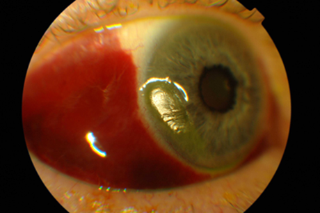
Figure 1: Right corneal dellen.
On exam, no facial injuries were noted, although there was some right upper lid swelling. Visual acuity on Snellen was 6/18 improving to 6/9 with pin hole right eye and 6/9 improving to 6/6 with pin hole left eye. Left ocular exam was generally unremarkable, aside from a few drusen noted on dilated fundal exam. There was no relative afferent pupillary defect (RAPD), ocular movements were full and painless, there was no diplopia and fields to confrontation, although difficult to assess with the right eye, appeared full. Isihara was full in both eyes. There was right lid erythema, no laceration evident and no periorbital swelling. There was extensive right subconjunctival haemorrhage and an inferior corneal dellen, approximately 2mm x 0.3mm (Figure 1). There was the odd cell present in the anterior chamber although the vitreous was quiet. Examination of the lens revealed non significant cataract and the intraocular pressure was normal at 12mmHg. Dilated right fundal exam revealed a few drusen only; there was no haemorrhage, no commotio and no tear or break evident.
Immediate management was commenced; two hourly lubricants (Hylofort tears) and four hourly Oc Chloramphenicol 1%. A follow-up review was planned for 24 hours later.
At next review, visual acuity on Snellen was unchanged at 6/18 improving to 6/9 with pin hole in the right eye, although the patient symptomatically felt better. The subconjunctival haemorrhage was still extensive and corneal dellen still evident. It was decided to continue with the same treatment plan and to review the patient again in five days.
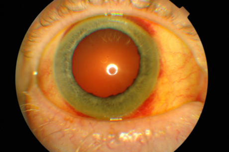
Figure 2: Resolved corneal dellen.
At second follow-up, our patient felt that aside from some residual redness, his eye was completely “back to normal”. Vision was 6/9 unaided in the right eye and on corneal exam, the corneal dellen had completely resolved (Figure 2). The patient was advised to stop using Oc Chloramphenicol 1% and to continue lubricants (Hylofort tears) four times a day. A final review was planned for him in six weeks.
At final review, our patient’s right visual acuity was stable at 6/9 unaided and his subconjunctival haemorrhage had completely resolved and his cornea was clear. Our patient was discharged with the ocular pathology completely resolved.
Discussion
This case illustrates that ocular trauma can result in acute corneal dellen formation. We postulate that the mechanism of acute onset of corneal dellen formation in this case was the ocular trauma sustained when our patient was injured with a foreign body whilst grass strimming. It is interesting to discover that corneal dellen can form so quicky and this may have an impact on the future management of this condition; it is well documented that if left untreated, or treated suboptimally, underlying corneal stroma may undergo secondary degeneration, leading to scarring and vascularisation and consequentially, loss of acuity [8]. Gradual development of corneal dellen, in patients already under the care of an ophthalmology team, can be managed appropriately with good outcome.
References
1. Tessler HH, Urist MJ. Corneal dellen in the limbal approach to rectus muscle surgery.
Brit J Ophth 1975;59:377-9.
2. Mai G, Yang S. Relationship between corneal
dellen and tearfilm breakup time.
Yan Ke Xue Bao 1991;7(1):43-6.
3. Fresina M, Campos E. Corneal “dellen” as a complication of strabismus surgery.
Eye (Lond) 2009;23(1):161-3.
4. Jampel HD, Musch DC, Gillespie BW, et al. Perioperative complications of trabeculectomy
in the Collaborative Initial Glaucoma Treatment Study (CIGTS). Am J Ophthalmol 2005;140:16-22.
5. Wood TO, Williams EE, Hamilton DL, Williams BL. Pterygium surgery with mitomycin and tarsorrhaphy. Trans Am Ophthalmol Soc 2005;103:108-15.
6. Ng LH. Central corneal epitheliopathy in a long term, overnight orthokeratology lens wearer: a case report. Optom Vis Sci 2006;83(10):709-14.
7. Moesen I, Hafezi F, Paridaens D. Corneal dellen secondary to conjunctival chemosis following transconjunctival orbital decompression. Klin Monbl Augenheilkd 2007;224(11): 856-7.
8. Dua HS, Forrester JV. Clinical patterns of corneal epithelial wound healing. Am J Ophthalmol 1987;104:481-9.
COMMENTS ARE WELCOME


