Blunt orbital trauma is a common consequence of sports injuries. Although retrobulbar haemorrhage is frequently associated with orbital compartment syndrome (OCS), the severity of clinical signs can outweigh imaging findings.
Early recognition and intervention, such as lateral canthotomy and cantholysis (LCC), are essential to prevent irreversible vision loss. This case explores the management of a young ENT surgeon who sustained orbital trauma during a football match.
Case Report
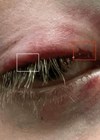
A 30-year-old female ENT surgeon presented to the emergency department (ED) after sustaining blunt orbital trauma during a football match. The injury occurred when the ball deflected off another player and directly struck her left eye (LE).

Figure 1a: CT facial bones, brain, sinuses without contrast – thick, soft.

Figure 1b: CT facial bones, brain, sinuses without contrast – thick, bones.

Figure 2a: CT facial bones, brain, sinuses without contrast – thin, soft.

Figure 2b: CT facial bones, brain, sinuses without contrast – thin, bones.

Figure 3a–d: Lateral canthotomy.
On initial assessment, the patient exhibited significant periorbital swelling in the LE, a rapid afferent pupillary defect (RAPD), corneal oedema, and immediate vision loss. She could only perceive colours with the affected eye. Intraocular pressure (IOP), measured using a handheld tonometer, was elevated to 43mmHg in the LE, while the right eye measured 18mmHg. The use of the handheld tonometer represented a limitation, as the gold standard, Goldmann applanation tonometer (GAT), was unavailable for use correctly overnight in the ED [1,2]. The patient was visibly anxious and expressed concern about her vision. Essentially, the patient’s career was pending on this moment as the loss of vision in her affected eye would have led to the loss of stereoacuity and depth perception, impacting her ability to train as a surgeon.
Computed tomography (CT) imaging showed no acute intracranial abnormality with minimal periorbital haemorrhage without evidence of significant optic nerve compression. Despite this, clinical findings strongly indicated OCS, prompting the decision to proceed with an emergency LCC.
The procedure was performed at the bedside, following local guidelines and using instruments from the HALO trolley (high acuity low occurrence). The steps involved included: local anaesthesia was administered at the lateral canthus, followed by a 1–2cm incision at the lateral canthal tendon using curved scissors. The inferior limb of the tendon was then cut to release orbital pressure [3]. This resulted in an immediate return of vision in the LE and a significant reduction in orbital tension. Her vision in the LE before transfer to the tertiary centre had returned to 6/12.
The patient was subsequently taken under the care of ophthalmologists for further evaluation and management. She was discharged the next day with no residual visual deficits and 6/6 vision bilaterally. The patient was advised to apply topical antibiotic ointment to reduce the risk of infection and to use cold compresses to minimise swelling and bruising. After discharge from the ophthalmology team, follow-up was arranged within 24 hours to assess healing and ensure there were no secondary complications, such as ongoing OCS. She was instructed to avoid excessive eye rubbing or pressure on the affected area. Regular visual acuity and IOP monitoring were emphasised to detect any delayed issues. If necessary, suture repair could be performed later to address any cosmetic concerns, though this was not considered urgent at the time of discharge.
Discussion
This case highlights the importance of prioritising clinical assessment over imaging in cases of suspected OCS. Retrobulbar haemorrhage may appear minimal on imaging but can still result in elevated orbital pressure, compromising optic nerve perfusion. This discrepancy arises because CT scans primarily detect high-density haematomas but may not adequately reflect the dynamic pressure effects of evolving OCS. The retrobulbar space is relatively enclosed, meaning that even small volumes of haemorrhage can significantly increase pressure, leading to optic nerve ischaemia and functional impairment. Other factors also include significant orbital oedema, and severe inflammation [4].
Key clinical markers – such as RAPD, vision loss, corneal oedema, and elevated IOP – are critical in guiding timely intervention. Handheld tonometry, while limited compared to GAT, provided a rapid, actionable IOP measurement that supported the diagnosis [5]. Early recognition by the triage nurse and medical team was key to ensuring treatment was received within one hour.

Figure 4a–c.
High acuity, low occurrence procedures include lateral canthotomy, subclavian access, resuscitative hysterotomy, emergency delivery, and emergency amputation. The HALO trolley (Figures 4a–c) ensures that the necessary equipment for these rare but life-saving interventions is easily accessible. Step-by-step guides are included to assist clinicians in executing these procedures efficiently in high-pressure scenarios.
Lateral canthotomy and cantholysis are low-risk, sight-saving procedures that should not be delayed in the context of OCS. Immediate intervention can lead to dramatic improvement, as demonstrated in this case, where vision returned immediately post-procedure. Delays in treatment due to reliance on imaging alone may result in irreversible optic nerve damage. Finally, this case emphasises the value of clinician intuition, or ‘gut feeling’, in recognising early signs of deterioration, even when imaging findings are equivocal.
References
1. Cook JA, Botello AP, Elders A, et al. Systematic Review of the Agreement of Tonometers with Goldmann Applanation Tonometry. Ophthalmology 2012;119(8):1552–7.
2. Stamper RL. A history of intraocular pressure and its measurement. Optom Vis Sci 2011;88(1):E16–28.
3. Amer E, Abbas AER. Ocular compartment syndrome and lateral canthotomy procedure. J Emerg Med 2019;56(3):294–7.
4. Borgenvik H, Ramsay MW, Nilsson M. Comparison between non-contact and rebound tonometry in patients with acute eye symptoms. Int J Ophthalmic Prac 2011;2(3):111–6.
5. Murali S, Davis C, McCrea MJ, Plewa MC. Orbital compartment syndrome: Pearls and pitfalls for the emergency physician. J Am Coll Emerg Physicians Open 2021;2(2):e12372.
Declaration of competing interests: None declared.