An experienced ophthalmologist can make an anatomical diagnosis of childhood visual impairment based upon the surgical sieve, i.e., congenital and acquired. But an ophthalmologist cannot work in isolation to make an aetiological diagnosis – one would require the help of a team consisting of paediatricians, neurologists, radiologists and geneticists. This is the case report of a child where the anatomical diagnosis was made with the first presentation, but a definite aetiological diagnosis was made much later with the help of a molecular genetic team.
Case report
A seven-month-old baby girl was referred to the clinic with concerns from her Paediatrician that the baby was not fixing or following. The baby was born two weeks premature, had a normal birth weight, uneventful pregnancy, uneventful delivery and healthy neonatal period. There was no positive family history of any eye disease.
The orthoptist and ophthalmologist who examined the baby’s eyes in the outpatient clinic found no abnormal ocular motility, nystagmus or relative afferent pupillary defect. However, the eyes did not fix and follow light. She had normal ocular anterior segments with no cataract. The ocular fundi were normal except for bilateral excavation of the optic disc with very thin neuro-retinal rim. The fundus photographs were taken when she was much older (Figures 1 and 2).
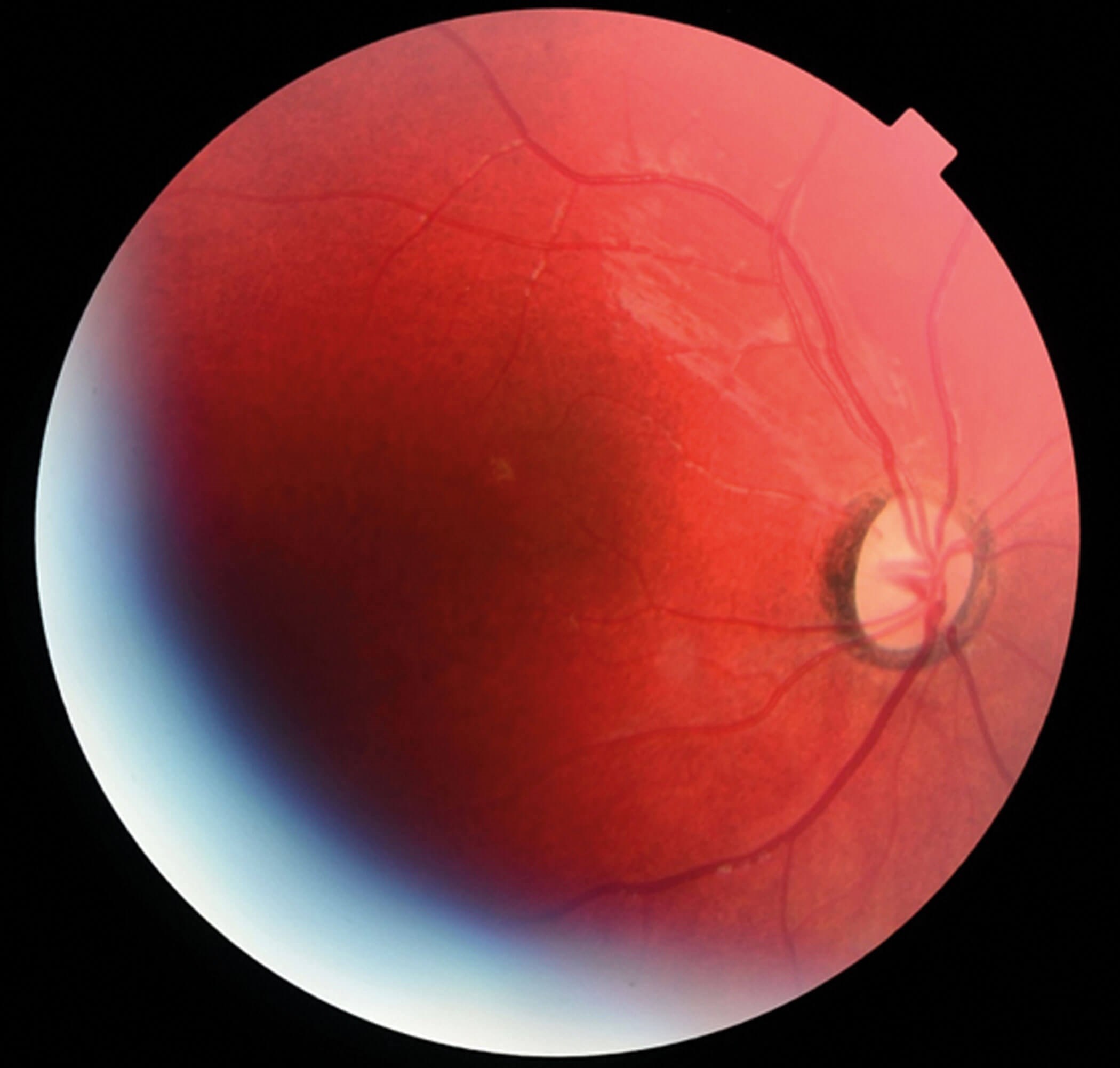
Figure 1: Right ocular fundus.
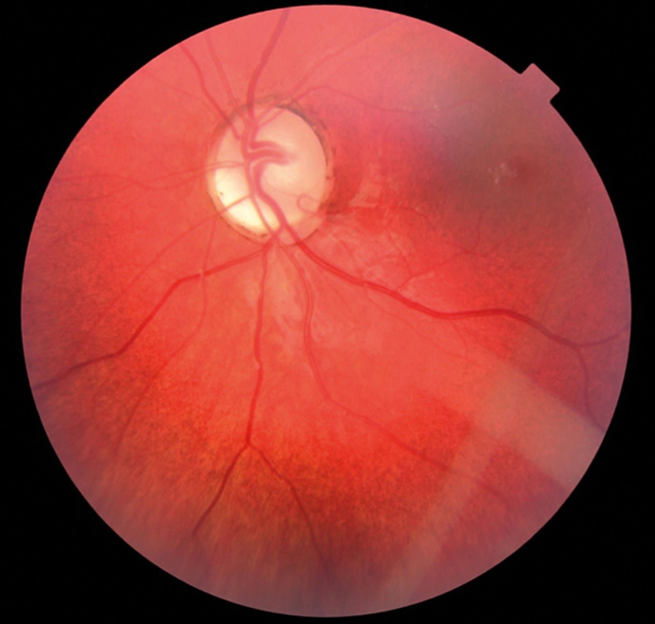
Figure 2: Left ocular fundus showing excavated optic discs.
An initial diagnosis of bilateral congenital optic disc hypoplasia was made pending further investigations. Magnetic resonance imaging (MRI) under general anaesthesia showed bilateral small size optic nerves, but no other intracranial abnormality. Electro-diagnostic tests (EDT) revealed poor primary visual pathway response.
This child continued to be followed up with the help of the hospital multidisciplinary team. At her current age of eight years, she is orally fed and has global developmental delay. Being wheelchair bound, she lacks the physical strength to stand up unaided and can only move independently when sitting on the floor and shuffling. Her speech has continued to improve and she can understand simple instructions. She has been attending a special needs school. The multidisciplinary team included a physiotherapist, speech and language therapist, occupational therapists and physician. The child is registered with severe visual impairment.
At her most recent clinic visit, her visual status had improved slightly and she was able to fix and follow light with both eyes open. No significant refractive error was found since referral.
The child was investigated by the regional genetic centre making the diagnosis of NR2F1 mutation causing optic atrophy and intellectual disability.
Discussion
Optic neuropathy is a frequent cause of visual impairment in children. Sometimes it can be a diagnostic challenge. Antenatal, perinatal, postnatal and family history can indicate a probable diagnosis in two thirds of children [1]. The initial diagnosis can be made by examination in the outpatient clinic or examination under general anaesthesia. Ancillary tests may identify the cause in the remaining cases. MRI and EDT are essential diagnostic tools along with molecular genetic testing to find out the underlying cause.
Bosch-Boonstro-Schaaf optic atrophy syndrome (BBSOAS) is a rare congenital neurodevelopmental disorder and has the course of static encephalopathy. It is an autosomal dominant disease. The majority of cases are de novo. It is caused by missense variants or deletions in the NR2F1 gene [2]. This is an important regulator of transcription in neurons and other tissues.
Patients show a variable degree of clinical features [2], such as:
- Ocular features: Optic atrophy, visual impairment, nystagmus, strabismus, alacrima.
- Systemic features: (General) global developmental delay, intellectual disorder, abnormal hearing, speech delay; (behavioural) cognitive anomalies, autism spectrum disorder; and (neuromuscular) general hypotonia, epileptic disorders, cranial anomalies.
On the positive side, these children love music and have good long-term memory and pain tolerance. There is also no major limitation in life expectancy.
Currently there is no curative treatment. However, with early intervention and supportive treatment much can be done to improve the skills and quality of life of these affected individuals. The recommendation is to involve early on a clinical geneticist in all cases of congenital eye diseases.
References
1. Jones R, Al-Hayouti H, Oladiwura D, et al. Optic atrophy in children: current causes and diagnostic approach. Eur J Ophthalmology 2020;30(6):1499-505.
2. Bosch DGM, Boonstra FN, Schaaf CP, et al. NRF2F1 Mutations cause optic atrophy with intellectual disability. Am J Hum Genet 2014;94(2):303-9.
Declaration of competing interests: None declared.
COMMENTS ARE WELCOME


