“It cost me an arm and a leg.” – Mr B told me. An arm and a leg to be seen by the famous Russian eye surgeon who said that everybody can be spectacle-free. He took Mr B’s money (roughly the equivalent worth of an arm and leg) and then said, well everybody except him and there is nothing that he could do about Mr B’s eyes and off he sent him from his ship. Researching a bit into this story, I think he spoke of Svyatoslav Fyodorov, the pioneer of radial keratotomy, which was performed in the famous assembly-line like rotating operating tables setting (Figure 1). Apparently, Professor Fyodorov also bought a vessel, the ‘Peter I’, and converted it into a floating eye clinic including theatres and cruised the oceans to come to meet his patients more than halfway.

Figure 1: The Moscow monument of Svyatoslav Fyodorov, the pioneer of radial keratomy, whom our
patient met on board his floating eye clinic. Picture from World Ophthalmology 2012;3(9):6.
Mr B, despite his eye problems, was in the navy (in a desk job to be fair though), when he climbed on board the Peter I at a time when his own ship was close by somewhere in the Mediterranean Sea in the 1980s. When Mr B presented three decades later to the former writer of this column and now Vitreoretinal Consultant, Mr Jon Park, the latter felt that the Professor’s promise may after all still apply to our patient.
Mr B went to a ‘blind school’ for his poor eyesight and it was not until later in his life that an optician figured out that with a very strong plus prescription he could actually see not too badly. The strong prescription was necessary because his crystalline lenses both fully dislocated making him aphakic in both eyes. Apparently, in the 1980s, Moorfields Eye Hospital investigated him for possible Marfan’s but no records prevailed and Mr B did not have any other symptoms indicative of that or another systemic condition. Indeed, he was generally fit and healthy when he went to see his optician last year for a foreign body he noted in front of his right iris (Figure 2). His corrected visual acuity with his heavy glasses (he did not tolerate contact lenses) measured 6/12 and 6/9 right and left, respectively.
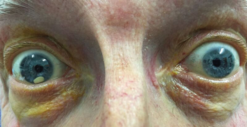
Figure 2: Dislocated lens fragment in the anterior chamber at presentation.
On the slit-lamp, this foreign body turned out to be his white lens still surrounded by capsule and another lens fragment was seen in the vitreous. In his left eye, his lens was also found to be dislocated, here fully into the vitreous. He soon underwent right lens fragment removal from the anterior chamber with a vectis loop combined with vitreo-lensectomy to remove residual fragments from the vitreous cavity. At the same sitting, an anterior chamber intraocular lense (IOL) was implanted (with a peripheral iridotomy).
Discussion
Anterior dislocation of the crystalline lens can result in complications such as corneal oedema, acute glaucoma or uveitis. Unlike a lens dislocated into the vitreous cavity, an anteriorly dislocated lens should always be removed because of possible devastating complications from endothelial cell loss or secondary glaucoma [1]. In most cases trauma is the cause of the dislocation, but other causes are possible as well (Table 1).

All patients with non-traumatic lens dislocation without a known cause should undergo biochemical screening for Homocystinuria as thromboembolic events are common with this condition and may cause death at an early age [2].
To our knowledge this is the first described case of a spontaneous subsequent lens dislocation from the vitreous where it had previously dislocated into the anterior chamber, that is not associated with trauma or systemic disease. Only two case reports could be found that described the same phenomenon but this was in a trauma case and Marfan’s patient respectively [3]. As can be seen from Figure 2, a further unique feature of this case is that the crystalline lens seems to have split before starting to migrate as there was another piece still to be found in the vitreous cavity on presentation.
At his very recent review in clinics, six weeks after corneal suture removal, Mr B’s visual acuity with a very slight myopic correction measured 6/9 in the right eye and he felt his vision to be a “1000% better.” He had spent over 60 years being told he was effectively blind and struggled with aphakic glasses – so naturally he was delighted with the result and is eager to have a similar procedure for the fellow eye. And it didn’t even cost him an arm and a leg.
References
1. Jaffe NS, Jaffe MS, Jaffe GF: Cataract surgery and its complications. 6th ed. St Louis: Mosby; 1997.
2. Simon MA, Origlieri CA, Dinallo AM, et al. New management strategies for ectopia lentis. J Pediatr Ophthalmol Strabismus 2015;52(5):269-81.
3. Schäfer S, Spraul CW, Lang GK. Spontaneous dislocation to the anterior chamber of a lens luxated in the vitreous body: two cases. Klin Monbl Augenheilkd 2003;220(6):411-3.
Acknowledgments:
I am very grateful to the previous author of this column, Mr Jon Park, Consultant Ophthalmologist at Musgrove Park Hospital, Taunton, for pointing my attention to this case and for helping with this article. We would both like to thank the patient for allowing the publication of his story.
COMMENTS ARE WELCOME


