
Photoessay
A refractive surprise after vitrectomy and capsulectomy
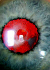
Figure 1: Right posterior capsule small aperture. We report a case of a pseudophakic patient who underwent vitrectomy and posterior capsulectomy. In spite of good visual acuity and absence of floaters, he was unhappy with the visual outcome. Case report...

Acute dellen formation post trauma
Corneal dellen are saucer-like thinnings, usually of the peripheral cornea [1]. Dellen formation is thought to be related to localised tear film instability [2], specifically the absence of the mucin component of the tear film. Without the mucin layer, dry...

A patient report of pseudoxanthoma elasticum, angioid streaks and choroidal neovascularisation
Angioid streaks (AS) on their own do not cause many problems, with the majority of patients remaining asymptomatic [1]. However, once choroidal neovascularisation (CNV) occurs, the visual prognosis of the patient rapidly declines [2]. Treatment is imperative to try and...

Acute retinal necrosis presumably caused by Epstein-Barr virus infection
Acute retinal necrosis (ARN) is an uncommon, but serious and potentially blinding condition. ARN is characterised by panuveitis, occlusive vasculopathy and progressive peripheral necrotising retinitis. The diagnosis is clinical but confirmation is sought via aqueous and vitreous sampling. Varicella zoster...

An unusual case of acute retinal necrosis
Case report A 40-year-old Caucasian male presented with a four-day history of redness and progressive painless reduction of vision in the left eye. His visual acuities were 6/4 in the right and 6/36 in the left. The left eye showed...

Severe conjunctival cicatrisation secondary to chronic glaucoma therapy
The timing of glaucoma filteration surgery during the course of chronic progressive glaucoma remains a contentious issue amongst glaucoma specialists. The vast majority support the use of maximal medical treatment initially to achieve the target pressure. Surgical procedures are only...

Melanocytoma of the optic disc
Melanocytoma is a heavily pigmented lesion, composed of melanocytes and is a variant of melanocytic naevus. It classically occurs at the optic disc and is usually benign, static and asymptomatic. However, it can occasionally grow and invade the retina or...

A single intravitreal bevacizumab in the treatment of breast carcinoma related choroidal metastasis
Case report Choroidal metastasis represents a common form of intraocular malignancies occurring in up to 10% of patients with systemic metastasis [1]. The most common primary sites of ocular metastasis are breast carcinoma in women and lung carcinoma in men,...
Birdshot chorioretinopathy: an important differential
Birdshot chorioretinopathy (BSCR) is a relatively uncommon cause of posterior uveitis which often has a relapsing and remitting course [1,2]. We present a case which demonstrates how remission can be obtained for several years using cyclosporine. Case report A 44-year-old...

Macula re-attachment following intravitreal ranibizumab in rhegmatogenous retinal detachment
Ranibizumab (Lucentis) is a vascular endothelial growth factor inhibitor (anti-VEGF) used for treatment of choroidal neovascular membrane [1]. We report a case where macula off inferior rhegmatogenous retinal detachment was misdiagnosed as wet age-related macular degeneration (AMD) and three intravitreal...

Ocular Dirofilariasis: a diagnosis on the rise?
Dirofilaria repens is one of 40 species of Dirofilaria. It is a zoonotic filarial nematode that commonly impacts dogs, however, human infection can occur through affected organs like skin, lung and eyes [1]. Although, there have been case reports of...

Cytomegalovirus retinitis post intravitreal triamcinolone in an immunocompetent patient with juvenile glaucoma
Figure 1: Fundus photograph shows active CMV retinitis four weeks after IVTA in the right eye. Figure 2: Fundus photograph of the left eye with no CMV retinitis; atrophic disc as a result of juvenile glaucoma. Case report A 29-year-old...

